If you are new to scleral lens fitting, toric peripheral curves (back surface toricity) may be confusing or challenging to evaluate.
First, it is important to note that toric peripheral curves are not the same as front surface toricity, which is used optically to correct for residual astigmatism. Toric peripheral curves provide landing zone alignment for non-spherical scleral surfaces. Dr. Greg DeNaeyer and colleagues found that only 6% of scleral shapes are actually spherical (1), yet there is still a large percentage of scleral fits that are using spherical peripheral curves/haptics.
Figure 1. A toric haptic design on a scleral lens.
What is toricity?
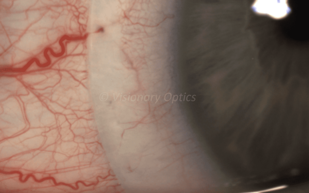
Simply put, toricity is the unequal curvature of two perpendicular meridians (Figure 1). When the scleral surface is more than 100 microns toric, then the landing zone of a scleral lens will benefit incorporating a toric design to properly align the ocular surface preventing compression and edge lift. The average scleral toricity at a 16 mm chord for the normal eye is 172 microns and for keratoconus with peripheral ectasia it’s 220 microns (2). A scleral lens with a spherical landing zone placed on a toric sclera will exhibit edge lift where the scleral surface is relatively steeper (Figure 2) and compression where the scleral surface is relatively flatter (Figure 3).
How do you fit scleral toricity?
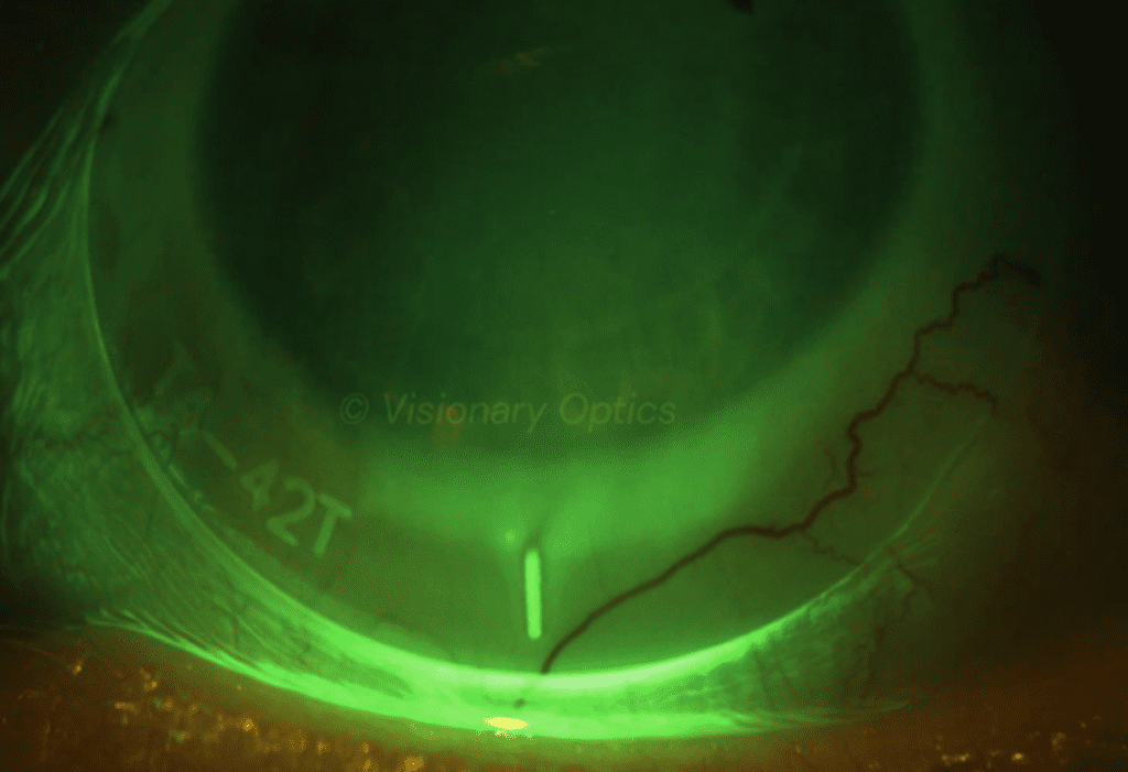
Europa and Europa Tangent fitting sets come with diagnostic lenses with spherical and toric landing zone designs. If a diagnostic lens with a spherical landing zone exhibits unequal areas of compression or edge lift, then apply another diagnostic lens with a 200 micron toric landing zone with similar sagittal depth. Hashmarks located at the periphery of the diagnostic lens indicate the steeper meridian. The lens will rotate so that the steeper lens meridian aligns with the steeper scleral meridian. It is important to note the position of the hashmarks in degrees or clock hours (Figure 4), as this might be needed as a reference when adding front surface toric correction for residual astigmatism correction or prism.
Next, evaluate the lens at the steep meridian and flat meridian 90 degrees away. Alignment of the landing zone without areas of edge lift or compression indicate the toricity of the lens properly matches the ocular surface. In the case of persistent misalignment, apply a diagnostic lens with 400 microns of back surface toricity with a similar sagittal depth and re-evaluate. Consultation can make any other fine tune adjustments in 100 microns steps if necessary.
What are the benefits of using toric peripheral curves?
Scleral lenses with toric peripheral curves provide improved landing zone alignment that results in better centration, comfort, and stability for the majority of fits. Individuals who struggle with fogging may find improvement in their vision as tear exchange will be reduced, limiting the rate at which debris build up in the tear reservoir (3-5).
Consultation is here to help
Communication with the consultants is critical to maximize your success. Make sure to let them know about any areas of residual misalignment that can be resolved with fine tune toric adjustments or in some cases a quadrant specific change.
The Visionary Optics team will help you provide the best fit for your patient!

Figure 3. An example of blanching of the conjunctiva due to a tight peripheral curve.
Figure 4. Europa Tangent Steep Meridian Hashmark.
- DeNaeyer G, Sanders D, van der Worp E, Jedlicka J, Michaud L, Morrison S. Qualitative Assessment of Scleral Shape Patterns Using a New Wide Field Ocular Surface Elevation Topographer: The SSSG Study. JCLRS [Internet]. 2017Nov.16 [cited 2023Jul.19];1(1):12-. Available from: http://www.jclrs.org/index.php/JCLRS/article/view/11
- DeNaeyer G, Sanders D, van der Worp E, Jedlicka J, Michaud L, Morrison S. Qualitative Assessment of Scleral Shape Patterns Using a New Wide Field Ocular Surface Elevation Topographer: The SSSG Study. JCLRS [Internet]. 2017Nov.16 [cited 2023Jul.23];1(1):12-. Available from: https://www.jclrs.org/index.php/JCLRS/article/view/11
- Jedlicka J, Johns L, Byrnes S. Scleral lens fitting guide. Contact lens spectrum 2010.Oct.1.Available from: https://www.clspectrum.com/issues/2010/october-2010/scleral-contact-lens-fitting-guide
- Johns, LK. Using Toricity with Scleral Lenses. Optometry Times. 2016Jun.17. Available from: https://www.optometrytimes.com/view/using-toricity-scleral-lenses
- Johns LK, McMahon J, Barnett M. Chapter 9 Scleral Lens Evaluation. Contemporary scleral lenses: theory and application. United Arab Emirates: Bentham Science Publishers, 2017: 199-240


