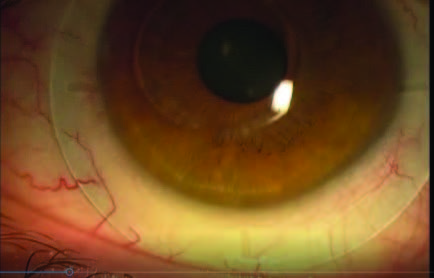
INDICATION: Keratoconus w/ Corneal Ring Implants
The novel topography device and innovative software technology accurately mapped the corneal and scleral irregularities, allowed Visionary Optics to empirically design a scleral lens with an “asymmetrically asymmetric” posterior haptic, and accurately predicted the fit and fluorescein patterns. This virtually fit scleral lens provided the patient with a successful scleral lens fit including good comfort and vision not experienced with previous lens designs.
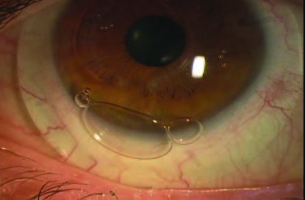
This patient had advanced keratoconus with corneal ring implants and habitually poor fitting scleral lenses resulting in a recurrent corneal ulcer overlying the bulging edge of one of the ring implants, and chronic lens intolerance. Patient had inferior lens lift and bubbles leaking under the lens (Figure 1).
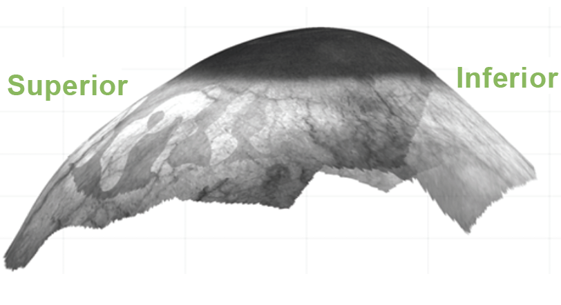
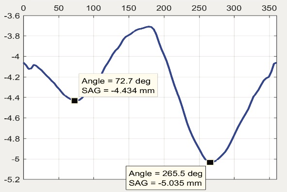
Qualitative 3D imaging with the sMap3D™ corneo-scleral topographer (Figure 2) demonstrated a much steeper scleral surface inferiorly than superiorly. A recent innovation with this instrument is the circumferential scleral shape plot which demonstrates the ocular SAG value (Y-axis) by axis meridian(X-axis) 360° around (Figure 3).
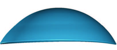
In this particular case, there was high scleral/conjunctival toricity, but with the steep axis inferiorly (266°) having a SAG >500μ greater than the steep axis superiorly (73°) [5035μ vs. 4434μ]. A custom lens was (Figure 4) was designed based on empirical corneo-scleral topography data to conform to this irregular scleral shape which had an optimal fit per OCT and slit lamp evaluation, was comfortable, provided excellent acuity and had no edge lift or bubble leakage (Figure 5).
Courtesy of Sheila Morrison OD, MS, FSLS
